Melanoma
The most serious and feared of all three forms of skin cancer is melanoma; Australia has the highest rate of melanoma in the world. Fortunately, Australia is also the most successful country when it comes to diagnosing and curing this type of cancer. If found within an appropriate margin; when less than 1mm deep, removing the cancer will cure 95% of people.
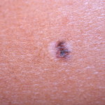
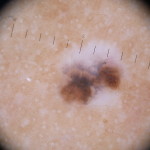
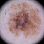
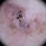
Most melanomas are brown and flat and appear on the skin as new spots; spots that haven’t always been there. However, a melanoma can be pink, bluish, black, white or a combination of colours, sometimes raised in appearance. They can also arise within an existing spot that has always been there. It is difficult to specify exactly what to look for as appearances of melanoma vary, but the most vital clue is watching for any change. Any new or existing spot which is changing shape or growing is suspicious and should be examined by a professional skin cancer clinic immediately. The majority of time, the spot is benign (non-cancerous), but the anxiety associated with a changing spot or mole can be enormous. To avoid feeling worried or anxious, please, make it a priority to have yourself checked.
In the case of a suspected melanoma, the affected spot will be surgically removed immediately and sent for pathology testing. If it is confirmed as a melanoma, the test results will inform us as to whether any further excision is necessary. This is done to notify us of the margin and ensure the melanoma has been thoroughly removed. Complete removal of the affected area is critical.
Once cured of melanoma, the next vital step is committing to regular, life-long check-ups. If you have had one melanoma you are at increased risk of getting another. Early diagnosis of any additional melanoma is crucial, ensuring that it too can be cured.
IF YOU HAVE EVER HAD A MELANOMA RING FOR AN APPOINTMENT


Basal Cell Carcinoma
A BCC, or Basal Cell Cancer, rises from the basal layer of the skins epidermis. This is the most common skin cancer and accounts for about ¾ of skin cancers treated in Australia. Unlike other forms of skin cancer, it does not shed cells which travel to distant sites in the blood or lymphatic vessels. However, it can still be a very serious skin cancer, particularly in certain areas of the body as it can grow down and damage important structures in the tissues beneath it.
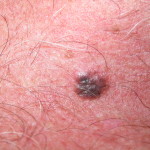
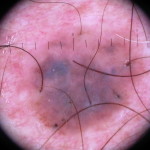
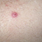
Most BCC’s occur on sun-exposed areas of the body such as the face and hands, although they can also occur on less exposed areas. Despite generally appearing as pink or skin-coloured, a BCC can occasionally be brown in colour. There are two main sorts of BCC’s: nodular and superficial. Nodular BCC’s appear as a small lump which is painless but occasionally ulcerates or bleeds. Superficial BCC’s look more like a flat, scaly, pink area. There are some rarer forms of Basal Cell Carcinoma that are much more serious and harder to diagnose. If you have previously had a BCC there is high chance you will get another, although this is the case with all forms of skin cancer. Anyone who has had any form of skin cancer should be diligent and regularly monitored and checked for the rest of their life. Early detection is the key to successful treatment with all forms of skin cancer.
IF YOU HAVE EVER HAD A BCC OR A SUSPICIOUS SPOT PLEASE RING FOR AN APPOINTMENT
Squamous Cell Carcinoma
Squamous Cell Carcinoma (SCC) is the second most common type of skin cancer. As the name implies, they arise from the Squamous layer of the skin which is found near the surface. Squamous Cell Carcinoma’s are more serious than BCC’s as they are able to easily invade the underlying tissues and can spread to lymph glands or distant organs. SCC’s occur most commonly on the head and neck, although legs and arms are also known to become affected. These cancers are generally caused by ultraviolet radiation (UVA and UVB) from the sun. Exposure to the much more carcinogenic UVC from welding can also be a cause. Without adequate protective clothing, 3 minutes of welding is thought to be the equivalent to 3 hours of sunbaking.
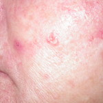
SCC’s present as a raised, scaly lump that may feel sore. They can appear on arms and legs resembling a mini-volcano; a cone with a slight depression in the centre. Sometimes there is also the presence a hard keratin core which can be picked off but will then reappear. SCC’s can grow in size at quite a rapid pace over a few days. Early forms may be flat and look a little like eczema. Aktinic keratosis (a sunspot) is the pre-malignant form of Squamous Cell Carcinomas. If left untreated, a significant proportion of these spots will transform into skin cancers. People with a large number of sunspots are at a much higher risk of becoming affected.
IF YOU HAVE EVER HAD A SCC OR A SUSPICIOUS SPOT PLEASE RING FOR AN APPOINTMENT
Sunspots
The technical term for Sunspots is solar keratoses or actinic keratoses. These are not a cancer but are pre-malignant, meaning they have the potential to turn into a squamous cell cancer. An affected sunspot may start as a small pink patch which will later develop a slight rough or scaly crust. Some may be hard to see and only detectable by their texture. There could be only a single affected spot, a few or perhaps several. This depends on an individual’s skin type, their amount of sun exposure and genetic heritage. The face is the most common area for sunspots to appear although they will occur anywhere on the body where the skin has experienced sun damage. Only a very small percentage of sunspots ever change into SCC’s but their purpose is to warn us that we have exposed the skin to a dangerous amount of ultraviolet rays and are therefore at risk of possibly developing skin cancer. Sunspots can be treated by applying facial creams and lotions and by method of freezing. Seek professional medical advice for information on sunspot removal.
Other Blemishes And Spots
There are many different types of skin lesions that can occur on anyone at any age. The following are benign and do not require treatment, (although if you are concerned, seek medical advice). Some of the most common are:
Seborrhoeic keratoses, age warts, age spots, senile warts, barnacles and liver spots: These are raised, dry or warty-looking spots. They can be flat, pale or dark. Sometimes, almost overnight they increase in size and can be the cause of great concern. Many people think these spots are unattractive and unsightly. This may be the case in some circumstances but never-the-less, they are harmless and not an indication of underlying skin cancer. Even though they are not a sign of old age, the older you become, the more you may develop. Spots such as these can either be ignored or frozen off.
Haemangiomas, aka Campbell de Morgan spots: These are bright red dots and can appear on any part of the body. They are generally the size of a pinhead although can sometimes be larger. They are caused by overgrowths of little blood capillaries, have no significance and are not dangerous. They can be ignored or removed.
Raised moles: Many moles are raised yet still perfectly normal. They are usually soft and floppy and easily traumatised by clothing or daily activities. It is possible to opt for their removal if they are noticeable, unattractive or causing discomfort.
Skin Tags: Skin tags are most commonly found in and around armpits and on the neck although can appear anywhere on the body. They range in size from very small to quite large and are skin-coloured or brown. Again, skin tags are harmless and removal is optional and straightforward.
Warts: Warts are not dangerous but are often difficult to treat. Many disappear over time if left alone.
Sebaceous hyperplasia: These spots usually occur on the forehead or other parts of the face. They are a small, pale nodule usually easy to diagnose with a dermatoscope and represent overgrown skin glands. They are of no particular significance nor are they of any concern.
Freckles, liver spots, brown spots, white spots: Changes to the skin caused by sun damage can result in flat brown spots on the back of hands or scaly brown or pink spots on the arms. The term “liver spots” can be misleading as no brown spot on the skin has any relevance to liver problems.
IF YOU HAVE EVER HAD A SUSPICIOUS SPOT PLEASE RING FOR AN APPOINTMENT